logo

EbookBell.com

Most ebook files are in PDF format, so you can easily read them using various software such as Foxit Reader or directly on the Google Chrome browser.
Some ebook files are released by publishers in other formats such as .awz, .mobi, .epub, .fb2, etc. You may need to install specific software to read these formats on mobile/PC, such as Calibre.

Please read the tutorial at this link:  https://ebookbell.com/faq 


We offer FREE conversion to the popular formats you request; however, this may take some time. Therefore, right after payment, please email us, and we will try to provide the service as quickly as possible.


For some exceptional file formats or broken links (if any), please refrain from opening any disputes. Instead, email us first, and we will try to assist within a maximum of 6 hours.

EbookBell Team
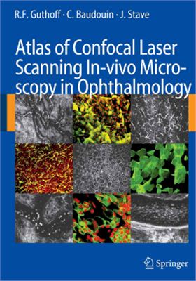
Atlas Of Confocal Laser Scanning Invivo Microscopy In Ophthalmology Guthoff Rf

  • SKU: BELL-5984004
Atlas Of Confocal Laser Scanning Invivo Microscopy In Ophthalmology Guthoff Rf
$ 31.00 $ 45.00 (-31%)

4.0

6 reviews

Atlas Of Confocal Laser Scanning Invivo Microscopy In Ophthalmology Guthoff Rf instant download after payment.

Publisher: Springer
File Extension: PDF
File size: 16.86 MB
Author: Guthoff R.F., Baudouin C., Stave J.
Language: English
Year: 2006

Product desciption

Atlas Of Confocal Laser Scanning Invivo Microscopy In Ophthalmology Guthoff Rf by Guthoff R.f., Baudouin C., Stave J. instant download after payment.

This atlas and textbook summarizes our experi-ence in the rather new field of clinical in vivo.
micromorphology. Stimulated by the work of Maurice, Jester, Bourne, Beuermann, Masters, Mathers, Thaer and others, our long-lasting cooperation is based on the mutual interest to push forward the frontiers of biomicroscopy using and combining recently available technical principles such as laser scanning, fast digital signal capture and processing and techniques for image stabilization. As a prerequisite there was the Heidelberg Retina Tomograph system.
for optic disc analysis with more than 5000 users worldwide and the engineering skill and.
dedication of physicists and engineers carefully listened to the wishes and dreams of the clinicians. So we are already on the way to in vivo biopsy, as postulated by Teruo Nishida.The content of the book reflects the common efforts of clinicians and physicists from the Rostock University Eye Department and the Centre Hospitalier National d’Ophtalmologie des Quinze-Vingts in Paris. Both groups share the enthusiasm to incorporate in vivo microscopy in the clinical decision making process by imag-ing, measuring and quantifying tissue parameters to achieve a quick and exact diagnosis and to monitor treatment in order to find the optimal path for patient recovery.
.
Contents:.
Introduction.
Principles of Confocal In Vivo Microscopy.
General Anatomical Considerations.
Ex Vivo Applications.
Confocal Laser Scanning In Vivo Microscopy.
Promising Applications.
Nonophthalmological Applications.
Confocal In Vivo Microscopy in Animal Experiments.
Current and Future Development.
References.
Subject Index.

Related Products