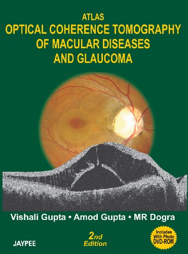
Product desciption
Atlas Optical Coherence Tomography Of Macular Diseases And Glaucoma 2nd Edition Vishali Gupta by Vishali Gupta, Amod Gupta, Mr Dogra 9788180616532, 8180616533 instant download after payment.
The emergence of optical coherence tomography (OCT) techniques has helped to look through the retina which actually is a 3- dimensional structure. The OCT provides a high resolution cross-sectional image of the macula very similar to the one obtained in vivo histopathological sections. It presents a major advance in the diagnostics of retinal disease. In this atlas the picture of stratus OCT (Tm) in various macular disorders helps to understand diagnosing and monitoring the response to various therapies and interventions and finding the correct therapeutic approach in a given patient. Its extensive application in diagnosis, management and follow-up of diabetic macular edema macular hole, taut posterior hyaloids membrane, vitreofoveal traction, idiopathic central serous chorioretinopathy, submacular pathology and many more areas. This atlas helps in easy comprehension because case summaries, fundus photographs, fluorescein angiography and the OCT images with followup program are provided about the patients. This is an adjunctive tool to probe the mysteries of retinal disease. This volume in ophthalmology presents emerging technologies that are shaping the world of refractive surgery. The readers will find an expert guidance on excimer laser instruments, wavefront technology, the recent advances made to photo ablation, refractive lens exchange and others. A concise format and numerous color photographs help the reader to hone the clinical skills while implementing the latest techniques. Plus a DVD featuring OCT images allows the reader to compare and choose the most appropriate machine for practice. · Features explanation of surgical techniques combined with high quality illustrations that emphasize and illuminate key points. · Includes pros and cons, pearls and pitfalls, and tricks of the trade for all techniques. · Presents detailed coverage of the main surgical areas in ophthalmic practice today. · Offers a consistent step-by-step approach to each procedure. · Lists equipment and instrumentation required for each procedures in appropriate conditions. · Uses full-color illustrations throughout the book. SECTION ONE : INTRODUCTION TO OCT, 1. Basics of Optical Coherence Tomography, 2. Technique of Acquiring OCT, 3. Selection of Scan Protocols, 4. Interpretation of OCT Scan SECTION TWO : OCT PATTERNS IN VARIOUS MACULAR DISEASES, 5. Diabetic Macular Edema, 6. Idiopathic Central Serous Chorioretinopathy (ICSC), 7. Macular Hole, 8. Retinal Vascular Occlusions, 9. Retinal Vasculitis, 10. Epiretinal Membranes, 11. Age-Related Macular Degeneration, 12. OCT in Choroidal Neovascular Membranes, 13. Juxtafoveal Telangiectasia, 14. Heredodystrophic Disorders, 15. Foveal Hemorrhage, 16. Photic Maculopathy, 17. Optic Disc Pits, 18. Inflammatory Diseases of Retina-choroid, 19. Retinal Angiomatosis Proliferation, 20. Retinal Trauma, 21. Macular Evaluation following Retinal Detachment Surgery, 22. Intraocular Tumors, 23. Degenerative Myopia, 24. Postoperative Endophthalmitis, SECTION THREE: GLAUCOMA, 25. Principles of OCT Scanning in Glaucoma, 26. The Normal Versus Glaucomatous Optic Disc, 27. OCT in the Clinical Management of Glaucoma, 28. Optical Coherence Tomography in Neuro-ophthalmology.