logo

EbookBell.com

Most ebook files are in PDF format, so you can easily read them using various software such as Foxit Reader or directly on the Google Chrome browser.
Some ebook files are released by publishers in other formats such as .awz, .mobi, .epub, .fb2, etc. You may need to install specific software to read these formats on mobile/PC, such as Calibre.

Please read the tutorial at this link:  https://ebookbell.com/faq 


We offer FREE conversion to the popular formats you request; however, this may take some time. Therefore, right after payment, please email us, and we will try to provide the service as quickly as possible.


For some exceptional file formats or broken links (if any), please refrain from opening any disputes. Instead, email us first, and we will try to assist within a maximum of 6 hours.

EbookBell Team
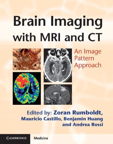
Brain Imaging With Mri And Ct An Image Pattern Approach 1st Edition Zoran Rumboldt Et Al Eds

  • SKU: BELL-5329384
Brain Imaging With Mri And Ct An Image Pattern Approach 1st Edition Zoran Rumboldt Et Al Eds
$ 31.00 $ 45.00 (-31%)

4.0

96 reviews

Brain Imaging With Mri And Ct An Image Pattern Approach 1st Edition Zoran Rumboldt Et Al Eds instant download after payment.

Publisher: Cambridge University Press
File Extension: PDF
File size: 48.35 MB
Pages: 428
Author: Zoran Rumboldt et al. (eds.)
ISBN: 9780521119443, 0521119448
Language: English
Year: 2012
Edition: 1

Product desciption

Brain Imaging With Mri And Ct An Image Pattern Approach 1st Edition Zoran Rumboldt Et Al Eds by Zoran Rumboldt Et Al. (eds.) 9780521119443, 0521119448 instant download after payment.

Most imaging books are ordered according to underlying etiology. However, in real life clinical practice, radiologists usually make their differential diagnoses according to the image patterns, as the etiology is often unknown. Brain Imaging with CT and MRI presents over 180 disease processes and normal variants, grouping entities by these basic patterns to accentuate differential diagnostic features. High quality CT and MRI scans show multiple typical and distinguishing images for each entity. Common and unusual clinical scenarios are described, including dilated perivascular spaces, capillary teleangiectasia, Susac's syndrome and desmoplastic infantile ganglioglioma. Both basic and advanced imaging techniques are used, reflecting the reality of clinical practice. This image-focused book emphasises the most pertinent clinical information relevant to the diagnostic process. Trainee and practising radiologists will find Brain Imaging with CT and MRI an invaluable and clinically relevant tool for learning and teaching.

Related Products