
Most ebook files are in PDF format, so you can easily read them using various software such as Foxit Reader or directly on the Google Chrome browser.
Some ebook files are released by publishers in other formats such as .awz, .mobi, .epub, .fb2, etc. You may need to install specific software to read these formats on mobile/PC, such as Calibre.
Please read the tutorial at this link: https://ebookbell.com/faq
We offer FREE conversion to the popular formats you request; however, this may take some time. Therefore, right after payment, please email us, and we will try to provide the service as quickly as possible.
For some exceptional file formats or broken links (if any), please refrain from opening any disputes. Instead, email us first, and we will try to assist within a maximum of 6 hours.
EbookBell Team

4.0
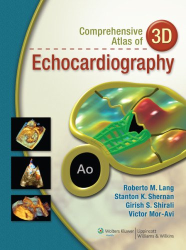
26 reviewsThe Comprehensive Atlas of 3D Echocardiography takes full advantage of today’s innovative multimedia technology. To help the reader understand the unique dynamic nature of a comprehensive 3D echocardiographic examination, the printed pages are supplemented with a companion website; this Atlas introduces the use of anatomy specimens, videos, unique imaging windows, and novel displays obtained with cropping tools. This approach offers a clear picture of how the diagnostic and monitoring capabilities of 3D echocardiography can benefit patients with a wide range of cardiovascular pathology, including congenital heart disease.
By showing a large number and variety of case studies, this Atlas demonstrates how 3D echocardiography can greatly enhance the diagnosis and clinical decision-making, especially when compared to two-dimensional techniques.
Whether you’re a Cardiologist, Sonographer, Anesthesiologist, Intensivist, Cardiac Surgeon, Researcher or any other Cardiovascular Medicine Professional, you’ll find this new Comprehensive Atlas of 3D Echocardiography is a must have reference book.
FEATURES
• Companion website includes more than 350 videos and 400 still images
• Emphasizes real-time 3D TEE technology
• Comparisons with anatomic specimens and 2D echocardiographic images provided, where helpful, to aid in the understanding of how 3D views and measurements relate to standard 2D techniques