logo

EbookBell.com

Most ebook files are in PDF format, so you can easily read them using various software such as Foxit Reader or directly on the Google Chrome browser.
Some ebook files are released by publishers in other formats such as .awz, .mobi, .epub, .fb2, etc. You may need to install specific software to read these formats on mobile/PC, such as Calibre.

Please read the tutorial at this link:  https://ebookbell.com/faq 


We offer FREE conversion to the popular formats you request; however, this may take some time. Therefore, right after payment, please email us, and we will try to provide the service as quickly as possible.


For some exceptional file formats or broken links (if any), please refrain from opening any disputes. Instead, email us first, and we will try to assist within a maximum of 6 hours.

EbookBell Team
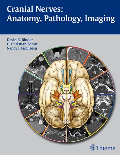
Cranial Nerves Anatomy Pathology Imaging 1st Devin Binder

  • SKU: BELL-2357948
Cranial Nerves Anatomy Pathology Imaging 1st Devin Binder
$ 31.00 $ 45.00 (-31%)

4.1

80 reviews

Cranial Nerves Anatomy Pathology Imaging 1st Devin Binder instant download after payment.

Publisher: Thieme
File Extension: PDF
File size: 137.13 MB
Pages: 248
Author: Devin Binder, D. Sonne, Nancy Fischbein
ISBN: 9781588904027, 1588904024
Language: English
Year: 2010
Edition: 1st

Product desciption

Cranial Nerves Anatomy Pathology Imaging 1st Devin Binder by Devin Binder, D. Sonne, Nancy Fischbein 9781588904027, 1588904024 instant download after payment.

This beautifully illustrated book combines a detailed exposition of the anatomy and function of the cranial nerves with practical coverage of clinical concepts for the assessment and differential diagnosis of cranial nerve dysfunction. An introductory chapter provides a brief overview of cranial nerve anatomy and function, skull base anatomy, classification of pathologies, and imaging approaches. Each of the twelve chapters that follow is devoted to in-depth coverage of a different cranial nerve. These chapters open with detailed discussion of the various functions of each nerve and normal anatomy. The authors then describe common lesions and present a series of cases that are complemented by CT images and MRIs to illustrate disease entities that result in cranial nerve dysfunction.Highlights:-Concise descriptions in a bulleted outline format enable rapid reading and review-Tables synthesize key information related to anatomy, function, pathology, and imaging-More than 300 high-quality illustrations and state-of-the-art CT and MR images demonstrate important anatomic concepts and pathologic findings-Pearls emphasize clinical information and key imaging findings for diagnosis and treatment-Appendices include detailed information on brainstem anatomy, pupil and eye movement control, parasympathetic ganglia, and cranial nerve reflexesThis book is an indispensable reference for practicing physicians and trainees in neurosurgery, neurology, neuroradiology, radiology, and otolaryngology-head and neck surgery. It will also serve as a valuable resource for students seeking to gain a solid understanding of the anatomy, function, and pathology of the cranial nerves.

Related Products