
Most ebook files are in PDF format, so you can easily read them using various software such as Foxit Reader or directly on the Google Chrome browser.
Some ebook files are released by publishers in other formats such as .awz, .mobi, .epub, .fb2, etc. You may need to install specific software to read these formats on mobile/PC, such as Calibre.
Please read the tutorial at this link: https://ebookbell.com/faq
We offer FREE conversion to the popular formats you request; however, this may take some time. Therefore, right after payment, please email us, and we will try to provide the service as quickly as possible.
For some exceptional file formats or broken links (if any), please refrain from opening any disputes. Instead, email us first, and we will try to assist within a maximum of 6 hours.
EbookBell Team

4.4
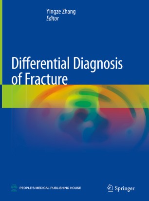
62 reviewsThis book covers diagnostic images of common and rare fractures for nearly every part of the human body, based on a large number of clinical cases. The highlight of this book is that both of three-dimensional X-ray images and CT/MRI images of thousands of fracture cases are presented for comparison and further discussion, according to the framework of AO classification.
The first chapter gives a general introduction of various diagnostic imaging techniques for fractures, with attention to their advantages and disadvantages. The following chapters present detailed radiological images of upper extremity fractures, lower extremity fractures, axial skeleton fractures, and epiphyseal lesions. It helps readers to recognize the difference between various diagnostic techniques, and to select optimal imaging techniques. With the illustrative figures, this book is a valuable tool to orthopaedist, radiologists, trauma surgeons, emergency room doctors, professional clinical staff, and medical students.