logo

EbookBell.com

Most ebook files are in PDF format, so you can easily read them using various software such as Foxit Reader or directly on the Google Chrome browser.
Some ebook files are released by publishers in other formats such as .awz, .mobi, .epub, .fb2, etc. You may need to install specific software to read these formats on mobile/PC, such as Calibre.

Please read the tutorial at this link:  https://ebookbell.com/faq 


We offer FREE conversion to the popular formats you request; however, this may take some time. Therefore, right after payment, please email us, and we will try to provide the service as quickly as possible.


For some exceptional file formats or broken links (if any), please refrain from opening any disputes. Instead, email us first, and we will try to assist within a maximum of 6 hours.

EbookBell Team
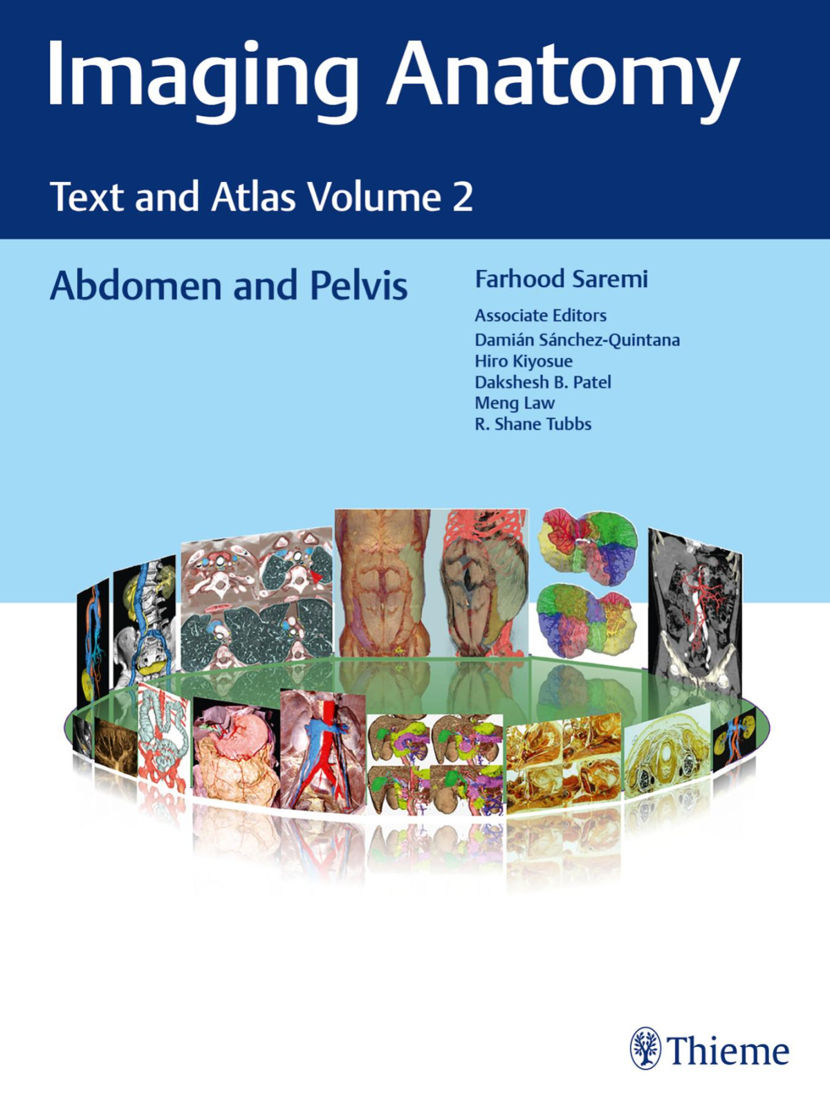
Imaging Anatomy Text And Atlas Volume 2 Abdomen And Pelvis 1st Farhood Saremi Editor In Chief

  • SKU: BELL-48191396
Imaging Anatomy Text And Atlas Volume 2 Abdomen And Pelvis 1st Farhood Saremi Editor In Chief
$ 31.00 $ 45.00 (-31%)

5.0

108 reviews

Imaging Anatomy Text And Atlas Volume 2 Abdomen And Pelvis 1st Farhood Saremi Editor In Chief instant download after payment.

Publisher: Thieme
File Extension: PDF
File size: 131.51 MB
Pages: 680
Author: Farhood Saremi (Editor in Chief)
Language: English
Year: 2023
Edition: 1st
Volume: 2

Product desciption

Imaging Anatomy Text And Atlas Volume 2 Abdomen And Pelvis 1st Farhood Saremi Editor In Chief by Farhood Saremi (editor In Chief) instant download after payment.

This imaging anatomy series is a reference atlas and text with
a practical discussion of specific topics along with examples of
anatomical variants. Divided into four major volumes, each volume
takes the instructive format of a classic text but is infused
with a large number of images and illustrations to teach readers
anatomy via state-of-the-art cross-sectional and volumetric
imaging. The volumes are divided into lung, mediastinum, and
heart; abdomen and pelvis; musculoskeletal; and head, neck,
and spine imaging. Peripheral vasculature and nerves have been
included in the musculoskeletal volume.
The length of each chapter and number of images vary in accordancewith the complexity of the topic. In all instances, effort has
been made to provide a concise yet comprehensive review of
the topic. Each chapter contains an in-depth review of the anatomy
and anatomical variants. Pertinent embryology, microanatomy,
and a brief description of physiology are discussed in each
chapter. Postsurgical anatomy and important gross and surgical
pathology images are included.
The text is supported by high-quality cross-sectional imageswith correlative three-dimensional and color-coded CT and MR
views. Up-to-date references are used to support the text. Many
new topics in radiology and surgery have been gathered from
the recent 10-year literature. Surgical and clinical applications in
each anatomy topic are presented with relevant images. In order
to make each topic understandable, difficult anatomical concepts
are supported by sketches as well as cross-sectional and topographic
cadaveric views provided by internationally known anatomists.

Related Products