Most ebook files are in PDF format, so you can easily read them using various software such as Foxit Reader or directly on the Google Chrome browser.
Some ebook files are released by publishers in other formats such as .awz, .mobi, .epub, .fb2, etc. You may need to install specific software to read these formats on mobile/PC, such as Calibre.
Please read the tutorial at this link: https://ebookbell.com/faq
We offer FREE conversion to the popular formats you request; however, this may take some time. Therefore, right after payment, please email us, and we will try to provide the service as quickly as possible.
For some exceptional file formats or broken links (if any), please refrain from opening any disputes. Instead, email us first, and we will try to assist within a maximum of 6 hours.
EbookBell Team
4.1
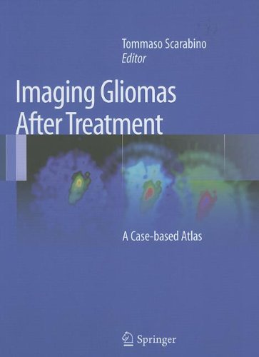
80 reviewsThis book illustrates the characteristics of imaging after treatment in brain gliomas. It describes in detail the modifications to brain tissue, both healthy and pathological, that can manifest after surgery, radiotherapy or chemotherapy treatment. These modifications are discussed in terms of both how they occur in the immediate post-treatment period, and in the long term. The imaging methods used include CT with and without the addition of contrast medium, but above all MRI, which involves the use of routine basic sequences and mainly advanced study techniques such as diffusion, perfusion, spectroscopy and cortical activation. The aim of the text is to equip neuroradiologists with adequate expertise in post-treatment examinations reporting, allowing them to perform an effective differential diagnosis between the persistency or recurrence of illness and the effects of short or long-term treatment.
The book is divided into a general section, which addresses the classification of cerebral tumors, the surgical treatment options, radiotherapy and chemotherapy protocols; and a section on clinical cases that employs rich iconography, making it quick and easy to consult.
This second edition has been updated to reflect the new WHO classification system from 2016; new surgical, radiotherapy and chemotherapeutic treatment options; and (in the iconography section) the new sequences available from the manufacturers of RM scanners.