Most ebook files are in PDF format, so you can easily read them using various software such as Foxit Reader or directly on the Google Chrome browser.
Some ebook files are released by publishers in other formats such as .awz, .mobi, .epub, .fb2, etc. You may need to install specific software to read these formats on mobile/PC, such as Calibre.
Please read the tutorial at this link: https://ebookbell.com/faq
We offer FREE conversion to the popular formats you request; however, this may take some time. Therefore, right after payment, please email us, and we will try to provide the service as quickly as possible.
For some exceptional file formats or broken links (if any), please refrain from opening any disputes. Instead, email us first, and we will try to assist within a maximum of 6 hours.
EbookBell Team
4.7
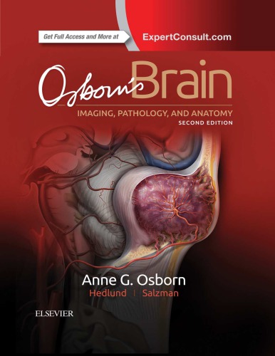
16 reviewsOsborn’s Brain: Imaging, Pathology, and Anatomy is the much-pleaded-for successor to Anne G. Osborn’s 1993 award winning book Diagnostic Neuroradiology (a.k.a. “The Red Book”), which became one of the all-time bestselling neuroradiology texts.
In this highly anticipated 1,200-page volume, Anne Osborn applies her special touch to make complex topics visually appealing and easy to understand. It wraps the “must know” aspects of brain imaging together with spectacular pathology examples, relevant anatomy, and the most up-to-date techniques in brain imaging.
Osborn’s Brain is organized for curriculum-based learning. Osborn begins with emergent topics, such as trauma, to help the reader learn in the order that is most practical for a resident or practicing radiologist. Indeed, this volume helps readers learn how to think about diagnoses, types of diagnoses, and the various pathologies that can affect the brain.
Even as Osborn “takes readers by the hand” to introduce them to the world of brain imaging, she includes new concepts and diagnoses that will intrigue the most sophisticated neuroradiologist.
The format of this book departs significantly from Osborn’s most recent publications. Rather than using bulleted text, she returns to detailed prose paragraphs designed to be a comprehensive learning resource. Summary boxes highlight the most important points from each section, enhancing functionality and ease of use.
This detail rich content is augmented with over 3,300 stunning, high resolution radiologic images and medical illustrations, all of which are annotated to describe the most clinically significant features.
Osborn’s Brain is easy to read, a visual delight, and a feast of concise, clear information. This impressive volume gives every radiologist and clinical neuroscientist exactly what he or she needs to know, while offering a fascinating journey into Osborn’s brain!