Most ebook files are in PDF format, so you can easily read them using various software such as Foxit Reader or directly on the Google Chrome browser.
Some ebook files are released by publishers in other formats such as .awz, .mobi, .epub, .fb2, etc. You may need to install specific software to read these formats on mobile/PC, such as Calibre.
Please read the tutorial at this link: https://ebookbell.com/faq
We offer FREE conversion to the popular formats you request; however, this may take some time. Therefore, right after payment, please email us, and we will try to provide the service as quickly as possible.
For some exceptional file formats or broken links (if any), please refrain from opening any disputes. Instead, email us first, and we will try to assist within a maximum of 6 hours.
EbookBell Team
5.0
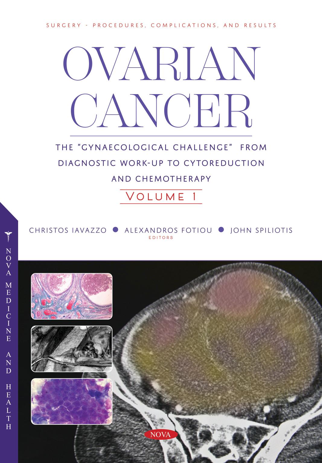
108 reviewsDiagnostic and pre-operative imaging has become increasingly adopted throughout the field of gynecology. In particular accurate preoperative analysis of ovarian pathology can improve the selection of the correct therapeutical approach, to reduce the length of operations, maximize surgical technique, and can ultimately improve a range of operative outcomes. New imaging modalities have advanced to the point of high resolution, three-dimensional analysis of tissue anatomy, composition and perfusion. Ultrasonography (US), Computed tomographic (CT) and magnetic resonance (MR) imaging have recently emerged as outstanding non-invasive techniques for the detection and characterization of ovarian pathology. In particular, US has probably now imposed itself as the “state-of-the-art” technique to explore the ovarian neoplasm, thanks the technical advancement like texture analysis (MGV) and 3D potentialities, although MR with new sequences like the diffusion-weighted imaging and greater magnetic fields (3 Tesla or more) may become leading methods in the future. The purpose of this book is to cover all the imaging techniques, potential for applying such imaging clinically, and to offer present and future applications as applied to ovarian pathology with the most world renowned scientists in these fields. The book is designed according to the pathological classification of the benign and malignant ovarian neoplasm by presenting for each pathology the clinical setting followed by the imaging approach. At the end of each chapter a “focus concept” paragraph will be presented with take-home point for the readers.