logo

EbookBell.com

Most ebook files are in PDF format, so you can easily read them using various software such as Foxit Reader or directly on the Google Chrome browser.
Some ebook files are released by publishers in other formats such as .awz, .mobi, .epub, .fb2, etc. You may need to install specific software to read these formats on mobile/PC, such as Calibre.

Please read the tutorial at this link:  https://ebookbell.com/faq 


We offer FREE conversion to the popular formats you request; however, this may take some time. Therefore, right after payment, please email us, and we will try to provide the service as quickly as possible.


For some exceptional file formats or broken links (if any), please refrain from opening any disputes. Instead, email us first, and we will try to assist within a maximum of 6 hours.

EbookBell Team
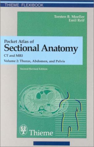
Pocket Atlas Of Sectional Anatomy Ct And Mri Volume 2 Thorax Abdomen And Pelvis 2nd Edition Torsten B Moller

  • SKU: BELL-873342
Pocket Atlas Of Sectional Anatomy Ct And Mri Volume 2 Thorax Abdomen And Pelvis 2nd Edition Torsten B Moller
$ 31.00 $ 45.00 (-31%)

0.0

0 reviews

Pocket Atlas Of Sectional Anatomy Ct And Mri Volume 2 Thorax Abdomen And Pelvis 2nd Edition Torsten B Moller instant download after payment.

Publisher: Thieme
File Extension: PDF
File size: 23.82 MB
Pages: 234
Author: Torsten B. Moller, Emil Reif
ISBN: 9780865778139, 9780865778726, 9783131255020, 9783131256027, 0865778132, 0865778728, 3131255021, 3131256028
Language: English
Year: 2001
Edition: 2
Volume: 2

Product desciption

Pocket Atlas Of Sectional Anatomy Ct And Mri Volume 2 Thorax Abdomen And Pelvis 2nd Edition Torsten B Moller by Torsten B. Moller, Emil Reif 9780865778139, 9780865778726, 9783131255020, 9783131256027, 0865778132, 0865778728, 3131255021, 3131256028 instant download after payment.

The second of a two-volume set which identifies the anatomical details visualized in the sectional imaging modalities. As a comprehensive reference, it is a great aid when interpreting images; schematic drawings of great clarity are juxtaposed with the CT and MRI images; anatomic structures are color-coded in the drawings to facilitate identification.

In this updated second edition, CT and MR images have been replaced with better quality substitutes.

Related Products