logo

EbookBell.com

Most ebook files are in PDF format, so you can easily read them using various software such as Foxit Reader or directly on the Google Chrome browser.
Some ebook files are released by publishers in other formats such as .awz, .mobi, .epub, .fb2, etc. You may need to install specific software to read these formats on mobile/PC, such as Calibre.

Please read the tutorial at this link:  https://ebookbell.com/faq 


We offer FREE conversion to the popular formats you request; however, this may take some time. Therefore, right after payment, please email us, and we will try to provide the service as quickly as possible.


For some exceptional file formats or broken links (if any), please refrain from opening any disputes. Instead, email us first, and we will try to assist within a maximum of 6 hours.

EbookBell Team
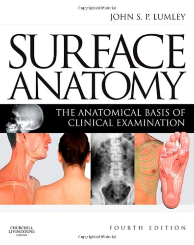
Surface Anatomy The Anatomical Basis Of Clinical Examination 4th John S P Lumley

  • SKU: BELL-7043186
Surface Anatomy The Anatomical Basis Of Clinical Examination 4th John S P Lumley
$ 31.00 $ 45.00 (-31%)

4.3

98 reviews

Surface Anatomy The Anatomical Basis Of Clinical Examination 4th John S P Lumley instant download after payment.

Publisher: Churchill Livingstone
File Extension: PDF
File size: 48.91 MB
Pages: 143
Author: John S. P. Lumley
ISBN: 9780443067945, 0443067945
Language: English
Year: 2008
Edition: 4th

Product desciption

Surface Anatomy The Anatomical Basis Of Clinical Examination 4th John S P Lumley by John S. P. Lumley 9780443067945, 0443067945 instant download after payment.

This book describes the visible and palpable anatomy that forms
the basis of clinical examination. It should be supplemented by
self-examination and the examination of normal subjects, and lead
to the examination of patients. Students and other models used
in practical sessions should come prepared with appropriate
clothing, such as swimwear, and there should be skin pencils
and tape measures to hand. In these sessions, a bony skeleton,
anatomical models and radiological material should be available
for comparison.
The first chapter considers the anatomical terms needed forprecise description of the parts of the body and movements
from the anatomical positions. There follows a description of
pathological terms, clinical examination and the additional tools
used in clinical practice.
The remaining chapters are regionally organised andcolour photographs demonstrate visible anatomy. Many of the
photographs are reproduced with numbered overlays, indicating
structures that can be seen, felt, moved or listened to. The surface
markings of deeper structures are indicated together with
common sites for injection of local anaesthetic, accessing blood
vessels, biopsying organs and making incisions. The text describes
the anatomical features of the illustrated structures.
The present edition has introduced figures to illustrate palpable
anatomy. Bony structures are green and deep bone green/brown.
Palpable arteries are red and palpable nerves yellow. Additional
muscle illustrations indicate the superficial subcutaneous muscles
encountered; these summary figures of muscle features can be
used as a quick reference and for revision purposes

Related Products