Most ebook files are in PDF format, so you can easily read them using various software such as Foxit Reader or directly on the Google Chrome browser.
Some ebook files are released by publishers in other formats such as .awz, .mobi, .epub, .fb2, etc. You may need to install specific software to read these formats on mobile/PC, such as Calibre.
Please read the tutorial at this link: https://ebookbell.com/faq
We offer FREE conversion to the popular formats you request; however, this may take some time. Therefore, right after payment, please email us, and we will try to provide the service as quickly as possible.
For some exceptional file formats or broken links (if any), please refrain from opening any disputes. Instead, email us first, and we will try to assist within a maximum of 6 hours.
EbookBell Team
4.3
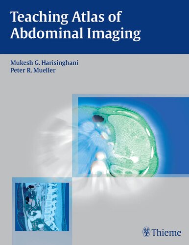
88 reviewsFeaturing 137 carefully selected cases, this atlas covers virtually every aspect of clinical cross-sectional imaging of the liver, gallbladder, biliary system and pancreas. For the vast majority of the cases, both CT and MR images are included to demonstrate the different features of each lesion. Furthermore, both typical and atypical pathologies are included to facilitate the differential diagnosis in daily clinical practice. Concise yet comprehensive,this atlas includes not only imaging features of the lesions but also the related pathologic and clinical data.
It is therefore useful both as a quick guide for practicing radiologists and as a brief textbook for radiologists in training.