Most ebook files are in PDF format, so you can easily read them using various software such as Foxit Reader or directly on the Google Chrome browser.
Some ebook files are released by publishers in other formats such as .awz, .mobi, .epub, .fb2, etc. You may need to install specific software to read these formats on mobile/PC, such as Calibre.
Please read the tutorial at this link: https://ebookbell.com/faq
We offer FREE conversion to the popular formats you request; however, this may take some time. Therefore, right after payment, please email us, and we will try to provide the service as quickly as possible.
For some exceptional file formats or broken links (if any), please refrain from opening any disputes. Instead, email us first, and we will try to assist within a maximum of 6 hours.
EbookBell Team
4.7
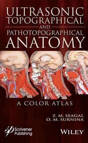
56 reviewsWritten by experienced and well-respected physicians and professors, this new all-color volume presents the ultrasonic topographical and pathotopographical anatomy of the body, including the head, neck, chest, anterolateral abdominal wall, abdominal organs, retroperitoneal space, male and female pelvises, and lower extremities.
Specific and non-specific ultrasonic symptoms are suggested for normal and abnormal developmental variants, diffuse and local pathotopographical anatomy. This color atlas contains comparative topographical and pathotopographical data and is the first manual of its kind for students and medical specialists in different areas, including those specializing in medical sonography. The original technology was tested at clinics in patients subjected to ultrasonic monitoring. Because of early detection there were no false-positive or false-negative results. The therapy was effective, and, in some cases, the use of the original method of “seagalography” (optometry and pulsemotorgraphy) has made it possible to develop new methods of treatment and/or to determine the optimal doses of drugs, as well as to develop effective drug complexes for treatment of a given pathology.
This important new volume will be valuable to physicians, junior physicians, medical residents, lecturers in medicine, and medical students alike, either as a textbook or as a reference. It is a must-have for any physician’s library.